Vermiform appendix
| Vermiform Appendix | |
|---|---|
 |
|
| Arteries of cecum and vermiform appendix. (Appendix visible at lower right, labeled as "vermiform process"). | |
 |
|
| Normal location of the appendix relative to other organs of the digestive system (frontal view). | |
| Latin | appendix vermiformis |
| Gray's | subject #249 1178 |
| System | Digestive |
| Artery | appendicular artery |
| Vein | appendicular vein |
| Precursor | Midgut |
| MeSH | Appendix |
| Dorlands/Elsevier | Vermiform appendix |
In human anatomy, the appendix (or vermiform appendix; also cecal (or caecal) appendix; also vermix) is a blind-ended tube connected to the cecum (or caecum), from which it develops embryologically. The cecum is a pouchlike structure of the colon. The appendix is located near the junction of the small intestine and the large intestine.
The term "vermiform" comes from Latin and means "worm-shaped".
Contents |
Size and location
The appendix averages 10 cm in length, but can range from 2 to 20 cm. The diameter of the appendix is usually between 7 and 8 mm. The longest appendix ever removed measured 26 cm from a patient in Zagreb, Croatia.[1] The appendix is located in the lower quadrant of the abdomen, or, more specifically, the right iliac fossa.[2] Its position within the abdomen corresponds to a point on the surface known as McBurney's point (see below). While the base of the appendix is at a fairly constant location, 2 cm below the ileocecal valve,[2] the location of the tip of the appendix can vary from being retrocecal (74%)[2] to being in the pelvis to being extraperitoneal. In rare individuals with situs inversus, the appendix may be located in the lower left side.
Vestigiality

The most common explanation for the appendix's existence in humans is that it's a vestigial structure which has lost its original function. (There has been little study of its function in the other animals in which it occurs—apes, wombats and some rodents—or comparison with animals in which it does not occur.) In The Story of Evolution, Joseph McCabe argued:
The vermiform appendage—in which some recent medical writers have vainly endeavoured to find a utility—is the shrunken remainder of a large and normal intestine of a remote ancestor. This interpretation would stand even if it were found to have a certain use in the human body. Vestigial organs are sometimes pressed into a secondary use when their original function has been lost.[3]
One potential ancestral purpose put forth by Charles Darwin[4] was that the appendix was used for digesting leaves as primates. It may be a vestigial organ, evolutionary baggage, of ancient humans that has degraded down to nearly nothing over the course of evolution. Evidence can be seen in herbivorous animals such as the koala. The cecum of the koala is very long, enabling it to host bacteria specific for cellulose breakdown. Human ancestors may have also relied upon this system and lived on a diet rich in foliage. As people began to eat more easily digested foods, they became less reliant on cellulose-rich plants for energy. The cecum became less necessary for digestion and mutations that previously had been deleterious were no longer selected against. These alleles became more frequent and the cecum continued to shrink. After thousands of years, the once-necessary cecum has degraded to what we see today, with the appendix.[4] On the other hand, evolutionary theorists have suggested that natural selection selects for larger appendices because smaller and thinner appendices would be more susceptible to inflammation and disease.[5]
Possible secondary functions
Immune function
New studies propose that the appendix may harbor and protect bacteria that are beneficial in the function of the human colon.[6]
Loren G. Martin, a professor of physiology at Oklahoma State University, argues that the appendix has a function in fetuses and adults.[7] Endocrine cells have been found in the appendix of 11-week-old fetuses that contribute to "biological control (homeostatic) mechanisms." In adults, Martin argues that the appendix acts as a lymphatic organ. The appendix is experimentally verified as being rich in infection-fighting lymphoid cells, suggesting that it might play a role in the immune system. Zahid[8] suggests that it plays a role in both manufacturing hormones in fetal development as well as functioning to "train" the immune system, exposing the body to antigens so that it can produce antibodies. He notes that doctors in the last decade have stopped removing the appendix during other surgical procedures as a routine precaution, because it can be successfully transplanted into the urinary tract to rebuild a sphincter muscle and reconstruct a functional bladder.
Maintaining gut flora
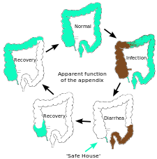
Although it was long accepted that the immune tissue, called gut associated lymphoid tissue, surrounding the appendix and elsewhere in the gut carries out a number of important functions, explanations were lacking for the distinctive shape of the appendix and its apparent lack of importance as judged by an absence of side-effects following appendectomy.[10] William Parker, Randy Bollinger, and colleagues at Duke University proposed that the appendix serves as a haven for useful bacteria when illness flushes those bacteria from the rest of the intestines.[6][11] This proposal is based on a new understanding of how the immune system supports the growth of beneficial intestinal bacteria,[12][13] in combination with many well-known features of the appendix, including its architecture and its association with copious amounts of immune tissue. Such a function is expected to be useful in a culture lacking modern sanitation and healthcare practice, where diarrhea may be prevalent.[11] Current epidemiological data[14] show that diarrhea is one of the leading causes of death in developing countries, indicating that as diarrhea flushes out the helpful bacteria the appendix helps recovery by providing a "safe house" for the bacteria.[11]
Diseases
The most common diseases of the appendix (in humans) are appendicitis and carcinoid tumors (appendiceal carcinoid).[15] Appendix cancer accounts for about 1 in 200 of all gastrointestinal malignancies. In rare cases, adenomas are also present.[16]
Appendicitis (or epityphlitis) is a condition characterized by inflammation of the appendix. Pain often begins in the center of the abdomen, corresponding to the appendix's development as part of the embryonic midgut. This pain is typically a dull, poorly localized, visceral pain.[17]
As the inflammation progresses, the pain begins to localize more clearly to the right lower quadrant, as the peritoneum becomes inflamed. This peritoneal inflammation, or peritonitis, results in rebound tenderness (pain upon removal of pressure rather than application of pressure). In particular, it presents at McBurney's point, 1/3 of the way along a line drawn from the anterior superior iliac spine to the umbilicus. Typically, point (skin) pain is not present until the parietal peritoneum is inflamed as well. Fever and an immune system response are also characteristic of appendicitis.[17]
Many cases of appendicitis require removal of the inflamed appendix, either by laparotomy or laparoscopy. Untreated, the appendix may rupture, leading to peritonitis, followed by shock, and, if still untreated, death.[17]
The surgical removal of the vermiform appendix is called an appendectomy, or appendicectomy.[18] This removal is normally performed as an emergency procedure when the patient is suffering from acute appendicitis. In the absence of surgical facilities, intravenous antibiotics are used to delay or avoid the onset of sepsis; it is now recognized that many cases will resolve when treated non-operatively. In some cases the appendicitis resolves completely; more often, an inflammatory mass forms around the appendix. This is a relative contraindication to surgery.
Use as efferent urinary conduit
The appendix is used for the construction of an efferent urinary conduit, in an operation known as the Mitrofanoff procedure,[19] in people with a neurogenic bladder.
Gallery

Mucinous adenocarcinoma of the appendix tip
|
See also
- Homology (biology)
- Noncoding DNA
References
- ↑ Guinness world record for longest appendix removed.
- ↑ 2.0 2.1 2.2 Paterson-Brown, S. (2007). "15. The acute abdomen and intestinal obstruction". In Parks, Rowan W.; Garden, O. James; Carter, David John; Bradbury, Andrew J.; Forsythe, John L. R.. Principles and practice of surgery (5th ed.). Edinburgh: Churchill Livingstone. ISBN 0-443-10157-4.
- ↑ McCabe, Joseph (1912). The Story of Evolution. London: Hutchinson & Co. http://onlinebooks.library.upenn.edu/webbin/gutbook/lookup?num=1043.
- ↑ 4.0 4.1 Darwin, Charles (1871) "Jim's Jesus". The Descent of Man, and Selection in Relation to Sex. John Murray: London.
- ↑ "The old curiosity shop". New Scientist. 2008-05-17. http://www.newscientist.com/channel/being-human/mg19826562.100-vestigial-organs-remnants-of-evolution.html.
- ↑ 6.0 6.1 Associated Press. "Scientists may have found appendix's purpose". MSNBC, 5 October 2007. Accessed 17 March 2009.
- ↑ "What is the function of the human appendix? Did it once have a purpose that has since been lost?". Scientific American. 1999-10-21. http://www.sciam.com/article.cfm?id=what-is-the-function-of-t. Retrieved 2008-07-01.
- ↑ Zahid, A. (April 2004). "The vermiform appendix: not a useless organ". J Coll Physicians Surg Pak 14 (4): 256–8. doi:04.2004/JCPSP.256258. PMID 15228837.
- ↑ Parashar, U.D.; Hummelman E.G.; Bresee J.S.; Miller M.A.; Glass R.I. (May 2003). "Global illness and deaths caused by rotavirus disease in children". Emerging Infect. Dis. 9 (5): 565–72. PMID 12737740. http://www.cdc.gov/ncidod/EID/vol9no5/02-0562.htm. "Table 1. Estimates of the annual number of diarrhea episodes among children <5 years of age in developing countries, by age group and setting".
- ↑ Kumar, Vinay; Robbins, Stanley L.; Cotran, Ramzi S. (1989). Robbins' pathologic basis of disease (4th ed.). Philadelphia: Saunders. pp. 902–3. ISBN 0-7216-2302-6.
- ↑ 11.0 11.1 11.2 Bollinger, R.R.; Barbas, A.S.; Bush, E.L.; Lin, S.S. & Parker. W. (21 December 2007). "Biofilms in the large bowel suggest an apparent function of the human vermiform appendix". Journal of Theoretical Biology 249 (4): 826–831. doi:10.1016/j.jtbi.2007.08.032. ISSN 0022-5193. PMID 17936308. http://www.sciencedirect.com/science?_ob=ArticleURL&_udi=B6WMD-4PKXBXY-2&_user=10&_coverDate=12%2F21%2F2007&_alid=994927353&_rdoc=1&_fmt=high&_orig=search&_cdi=6932&_sort=r&_docanchor=&view=c&_ct=1&_acct=C000050221&_version=1&_urlVersion=0&_userid=10&md5=037821a4aac437b2fc1706b2347f313e.
- ↑ Sonnenburg J.L., Angenent L.T., Gordon J.I. (June 2004). "Getting a grip on things: how do communities of bacterial symbionts become established in our intestine?". Nat. Immunol. 5 (6): 569–73. doi:10.1038/ni1079. PMID 15164016.
- ↑ Everett M.L., Palestrant D., Miller S.E., Bollinger R.R., Parker W. (2004). "Immune exclusion and immune inclusion: a new model of host-bacterial interactions in the gut". Clinical and Applied Immunology Reviews 5: 321–32.
- ↑ Statistics on the cause of death in developed countries collected by the World Health Organization in 2001 show that acute diarrhea is now the fourth leading cause of disease-related death in developing countries (data summarized by The Bill and Melinda Gates Foundation). Two of the other leading causes of death are expected to have exerted limited or no selection pressure on humans in the distant past because one (HIV-AIDS) only very recently emerged and another (ischaemic heart disease) primarily affects people in their post-reproductive years. Thus, acute diarrhea may have been one of the primary disease-related selection pressures on the human population in the past. (Lower respiratory tract infection (pneumonia) is the remaining of the top four leading causes of disease-related death in third world countries.)
- ↑ "Appendix disorders Symptoms, Diagnosis, Treatments and Causes". Wrongdiagnosis.com. http://www.wrongdiagnosis.com/a/appendix_disorders/intro.htm. Retrieved 2010-05-19.
- ↑ "Statics about Appendix disorder". Wrongdiagnosis.com. http://www.wrongdiagnosis.com/a/appendix_disorders/stats.htm. Retrieved 2010-05-19.
- ↑ 17.0 17.1 17.2 Miller R., Kenneth; Levine, Joseph (2002). Biology. Pentice Hall. pp. 92–98. ISBN 0-13-050730-X.
- ↑ "appendicectomy - definition of appendicectomy". Farlex Incorporation.
- ↑ Mingin G.C., Baskin L.S. (2003). "Surgical management of the neurogenic bladder and bowel". Int Braz J Urol 29 (1): 53–61. doi:10.1590/S1677-55382003000100012. PMID 15745470. http://www.brazjurol.com.br/january_february_2003/Baskin_ing_53_61.htm.
Further reading
- "The vestigiality of the human vermiform appendix: A Modern Reappraisal"—evolutionary biology argument that the appendix is vestigial
- Appendix May Actually Have a Purpose—2007 WebMD article
- Smith, H.F.; R.E. Fisher, M.L. Everett, A.D. Thomas, R. Randal Bollinger, and W. Parker (October 2009). "Comparative anatomy and phylogenetic distribution of the mammalian cecal appendix". Journal of Evolutionary Biology 22 (10): 1984–99. doi:10.1111/j.1420-9101.2009.01809.x. ISSN 1420-9101. PMID 19678866. http://www3.interscience.wiley.com/journal/122544996/abstract.
- Cho, Jinny. "Scientists refute Darwin's theory on appendix". The Chronicle (Duke University), August 27, 2009. (News article on the above journal article.)
External links
- SUNY Labs 37:12-0102—"Abdominal Cavity: The Cecum and the Vermiform Appendix"
- Appendicitis, most common disease of appendix.
|
|||||||||||||||||||||||||||||||||||||||||||||||||||||||
|
|||||||||||||||||||||||||||||||||||||||||||||||||||||||||||||
